
Geoduck clam (Panopea generosa): Anatomy, Histology, Development, Pathology, Parasites and Symbionts
Normal Histology - Organs Associated with the Visceral Mass
The body of the visceral mass contains the digestive, reproductive and excretory systems. In addition, the following organs are associated with the visceral mass:
Foot
The foot of most clams is a muscular organ directed towards the anterior end as an adaptation for burrowing. It is a muscular outgrowth of the visceral mass or body of the geoduck clam. In adult geoduck clams, the foot is relatively small in comparison to the body size. In juvenile geoduck clams the foot is comparatively large and is roughly equivalent in portion to the body size as in most other clams. The relatively large size of the foot in juveniles aids digging in the substrate whereas the small foot of adults accounts for their incapability of reorienting in the substrate once they have been disturbed. The foot can be extended through the pedal aperture (see drawing, Fig. 4 on Anatomy Page) or may be entirely withdrawn into the mantle cavity (as illustrated in histological sagittal sections on the Histology Overview Page). In juvenile clams of up to two years in age, a functional byssal gland is located in the proximal region at the posterior end of the foot (Fig.1a). This byssal gland produces byssal fibers (Figs.1b and 1c) which aid the geoduck in attaching to the substrate.

Figure 1a. The foot (f) is within the infrabranchial chamber (ib) of the mantle cavity and is extended between the labial palps (lp) located just posterior to the anterior adductor muscle (am). The byssal gland complex (b) is located at the base of the foot near the pedal aperture (pa) in the muscular mantle wall (mm).

Figure 1b. A longitudinal section through the base of the foot illustrating the byssal gland (b) and small ducts (bd) that join to the main canal containing byssal gland secretions (bf).

Figure 1c. A longitudinal section through the opening (o) of the byssal gland duct (bd) which contains byssal fibers (bf).
Figures 1a to 1c. Longitudinal sections through the visceral mass of juvenile geoduck clams. Haematoxylin and eosin stain.
Muscles
In addition to the anterior and posterior adductor muscles (labeled as am and pm, respectively, on Figs 1a and 1b of the Histology Overview Page), a paired band of muscle fibers pass through the left and right ventral regions of the kidney (Fig. 2). These muscle bands join and continue into the posterior curve of the visceral mass to the base of the foot, forming the retractor muscle of the foot.
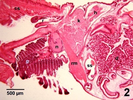
Figure 2. Histological section through the retractor muscle of the foot (rm) in the posterior region of a juvenile geoduck clam. This muscle passes between the style sac (ss) and the visceral (posterior) nerve ganglion (n) and divides into two bands that pass through the kidney (k). Other organs labeled on this figure include the gills (g), cut surface of the siphon (cs), rectum (r), heart (h), and digestive gland (d). Haematoxylin and eosin stain.
Nervous System
The geoduck clam has a typical bivalve nervous system containing three pairs of ganglia (Fig. 3a): cerebral ganglia on the left and right sides of the esophagus (Fig. 3b), a fused pair of pedal ganglia at the base of the foot within the visceral mass (Fig. 3c) and a pair of closely adjacent visceral (posterior) ganglia ventral to the posterior adductor muscle (Figs. 3d and 3e). The visceral ganglia are the largest of the three pairs and extend laterally through the body from the vicinity of the kidneys to the region of the posterior adductor muscle. In geoduck clams, there appears to be a trend towards centralising nervous coordination in the visceral ganglia as indicated by their larger size. Characteristic of mollusc ganglia, the nerve cell bodies are concentrated in the outer layer (cortex) and the nervous system is contained in a perineural sheath formed of glial cell elements (Morse and Zardus 1997).

Figure 3a. Longitudinal section showing the relative positions of the three pairs of ganglia: the cerebral ganglion (cg) anterior to the labial palps (lp) and ventral to the anterior portion of the digestive gland (dg), the pedal ganglion (pg) ventral to the intestine (i) at the base of the foot (f), and the visceral ganglion (vg) posterior to the kidney (k).
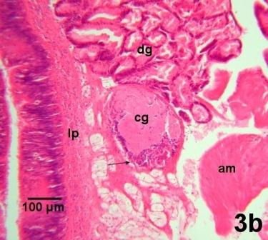
Figure 3b. Edge of one cerebral ganglion (cg) located between the anterior adductor muscle (am) and the labial palps (lp). The basophilic nerve cell bodies (arrow) are located ventral to the nerve fibers which are emerging from the cerebral ganglion towards the digestive gland (dg).

Figure 3c. A cross section through the fused pair of pedal ganglia (pg) with nerve fibers (arrows) extending towards the digestive gland (dg).

Figure 3d. Magnification of the visceral ganglion (vg) adjacent to the kidney (k). Other tissues in this section include the foot retractor muscle (rm), gills (g), edge of the anus (an), the heart (h) containing the rectum (rc), and the edge of the digestive gland (dg).

Figure 3e. A section through the visceral ganglion (vg) close to the posterior adductor muscle (pm) and the rectum (rc). In this section, the basophilic cortex of the nerve cell bodies of the visceral ganglion completely surrounds the eosinophilic central neurophil. An adjacent nerve fiber (arrow) is located next to the epithelial layer that separates the visceral mass from the suprabranchial chamber (sb) of the mantle cavity.
Figures 3a to 3e. Histological sections through the nervous system of juvenile geoduck clams. Haematoxylin and eosin stain.
A pair of statocysts are located anterior to the pedal ganglia. Although the statocysts are well developed in pediveliger larva (swimming stage late in larval development near the time of metamorphosis to benthic dwellers), they degenerate (Fig. 4) as the geoduck clam becomes established in the substrate.

Figure 4. Remnants of the statocyst (st) between nerve fibers (n) located at the base of the foot anterior to the pedal ganglia. Haematoxylin and eosin stain.
Hinge Complex
The hinge complex is a structure that lies on the dorsal surface of the visceral mass and produces the hinge ligament (Fig. 5).

Figure 5. Sagittal section through the dorsal area of a juvenile geoduck clam showing the hinge complex posterior and ventral to the hinge (hn). Haematoxylin and eosin stain.
References
Barnes, Robert D., 1968. Invertebrate Zoology, 2nd ed. W. B. Saunders Company, Toronto, 743 pp.
Morse, M.P. and Zardus, J.D. 1997. Bivalva. Microscopic Anatomy of Invertebrates Vol. 6A Mollusca II. F.W. Harrison and A.J. Kohn. Wiley-Liss. pp. 7-118.
Citation Information
Bower, S.M. and Blackbourn, J. (2003): Geoduck clam (Panopea generosa): Anatomy, Histology, Development, Pathology, Parasites and Symbionts: Normal Histology - Organs Associated with the Visceral Mass.
Date last revised: August 2020
- Date modified: