
Geoduck clam (Panopea generosa): Anatomy, Histology, Development, Pathology, Parasites and Symbionts
Normal Histology Digestive System
Foregut
Food particles that are gathered by the gills are moved to the labial palps which act as a device to sort and direct food particles to the mouth. Two pairs of labial palps, each pair located on either side of the body, occur at the anterior end of the geoduck clam on either side of the gills (see label "lp" in anatomy sketch and histological overview). The labial palps are mainly composed of vesicular connective tissue with basophilic gland cells (probably mucus secreting) within the epithelium along the inner margins. The inner surfaces are ridged and ciliated and act as a food particle sorting device (Fig. 1a and 1b). Food enters the mouth and is conducted through the tubular, ciliated esophagus to the stomach (Fig. 1c). The epithelial cells of the esophagus are larger, and have longer cilia than the epithelial cells lining the stomach (Fig. 1d).

Figure 1a. The labial palps located adjacent to the anterior nerve ganglion (ng) are mainly composed of vesicular connective tissue (vc) with an inner lining of ciliated epithelium (e) that forms ridges (arrowhead) towards the distal edges. Basophilic gland cells (gt) are located at the distal portion of the labial palps between the basement membrane (bm) of the epithelium and the vesicular connective tissue.

Figure 1b. A magnification of the epithelial ridges on the labial palps illustrates the transition between the muscle fibers (mf) and the glandular tissue (gt) below the basement membrane (bm) of the ciliated epithelium (ce).

Figure 1c. The anterior end of the alimentary canal showing the location of the mouth (m) between the labial palps (lp), the esophagus (e) and expanded stomach (s). The style sac (ss), digestive gland tubules (dgt), intestine (i), anterior nerve ganglion (ng), anterior adductor muscle (am) and foot (f) are also evident in this figure.

Figure 1d. A magnification of the junction between the esophagus and stomach illustrates the thicker epithelium and long cilia on the epithelial cells of the esophagus (e) in comparison to those of the stomach (s).
Figures 1a to 1d. Sections through the anterior end of the digestive tract of juvenile geoduck clams. Hematoxylin and eosin stain.
Midgut
The digestive gland surrounds the stomach and is composed of numerous tubules which are formed at the distal ends of branching ciliated ducts. Nutrients from the stomach enter the tubules through these ciliated ducts (Fig. 2a) and are absorbed by the glandular cells lining the tubules and digested intracellularly. The crystalline style extends from the style sac into the stomach (Fig.2b). The style contains amylase which helps digest the starches present in the food. In addition, the style usually (in other bivalves) harbours many spirochete bacteria that are thought to secrete additional digestive enzymes. These symbiotic bacteria have not yet been detected in Panopea abrupta. Wastes from the digestive gland tubules are returned to the stomach and are eventually swept into the intestine (Fig. 2c).
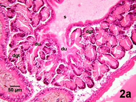
Figure 2a. Illustration of the ciliated ducts (du) leading from the stomach (s) to the digestive gland tubule (dgt).

Figure 2b. The eosinophilic crystalline style is within the style sac (ss) and extends into the stomach (s). Brown coloured excreta (ec) is evident at one end of the crystalline style and in adjacent sections through the intestine (i).
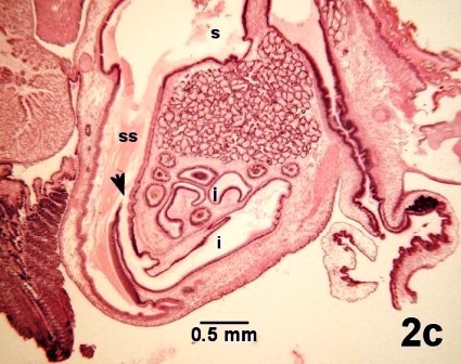
Figure 2c. Illustration of the opening of the duct (arrow) in the wall of the style sac (ss) that leads from the stomach (s) into the intestine (i).
Figures 2a to 2c. Sections through the digestive gland region of juvenile geoduck clams. Hematoxylin and eosin stain.
The excreta (wastes from digestion) proceed through the loops of intestine in the gonad (Fig. 3a to 3c). The wall of the intestine is composed of ciliated epithelium and mucus glands (Fig. 3d). The intestine continues dorsally and becomes the rectum.

Figure 3a. Intestinal loops (i) adjacent to the gonadal primordia (arrows) and digestive gland (dg) of a juvenile geoduck clam.
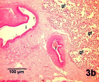
Figure 3b. Intestinal loops (i) adjacent to the gonadal follicles (gf) of developing ova in an adult female geoduck clam.

Figure 3c. Intestinal loops (i) adjacent to gonadal follicles (gf) containing spermatogonia and sperm in an adult male geoduck clam.

Figure 3d. A magnification of the intestinal wall consisting of ciliated epithelial cells (ce) and basophilic mucus gland cells (gt) and bordered by vesicular connective tissue (vc) of the clam and excreta (ec) within the lumen of the intestine.
Figures 3a to 3d. Sections through loops in the intestinal tract located between the digestive gland and gonad. Hematoxylin and eosin stain.
Hindgut
The rectum extends through the heart (Fig. 4a) and pericardial cavity (Fig. 4b) dorsal to the kidney and passes posterior to the posterior adductor muscle (Fig. 4c). The rectum opens through the anus at the posterior region of the suprabranchial space. The epithelial cells of the anus have elongate cilia (Fig. 4d).
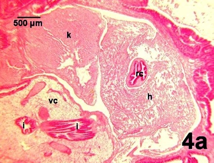
Figure 4a. The rectum (rc) is located within the heart (h) which is adjacent to the kidney (k). Sections through posterior regions of the intestine (i) occur near by in the vesicular connective tissue (vc).

Figure 4b. The rectum (rc) containing excreta (ec) is located in the pericardial space (pc) between lobes of the kidney (k) and the edge of the heart (h).

Figure 4c. The rectum (rc) passes posterior to the posterior adductor muscle (pm) and excreta within the rectum at the anus is located close to the visceral (posterior) neural ganglion (ng).

Figure 4d. A magnification of the anal region of Fig. 4c illustrates the elongate cilia of the ciliated epithelium (ce).
Figures 4a to 4d. Sections through the hindgut of juvenile geoduck clams. Hematoxylin and eosin stain.
As observed in the connective tissue of the labial palps (Fig. 1b above), glandular tissue also occurs in the connective tissue adjacent to the epithelial layer of the rectum at the anus (Figs. 5a and 5b).

Figure 5a. Arrows indicate patches of glandular tissue located in the connective tissue adjacent to the ciliated epithelium on the inner surface of the labial palps (lp) and the rectum (rc) at the anus. Other labeled organs in this section are: esophagus (e), stomach (s), digestive gland (dg), intestine (i), heart (h), kidney (k), posterior nerve ganglion (ng), foot retractor muscle (rm), gills (g), infrabranchial chamber (ib) and suprabranchial chamber (sb).

Figure 5b. A magnification of the glandular tissue (gt) in the connective tissue below the basement membrane (bm) of the ciliated epithelium (ce). The rectum is located towards the lower right hand corner of the figure.
Figures 5a and 5b. Sections through the glandular tissue associated with both ends of the digestive tract of juvenile geoduck clams. Hematoxylin and eosin stain.
References
Barnes, Robert D., 1968. Invertebrate Zoology, 2nd ed. W. B. Saunders Company, Toronto, 743 pp.
Morse, M.P. and Zardus, J.D. 1997. Bivalva. Microscopic Anatomy of Invertebrates Vol. 6A Mollusca II. F.W. Harrison and A.J. Kohn. Wiley-Liss. pp. 7-118.
Citation Information
Bower, S.M. and Blackbourn, J. (2003): Geoduck clam (Panopea generosa): Anatomy, Histology, Development, Pathology, Parasites and Symbionts: Normal Histology - Digestive System.
Date last revised: August 2020
- Date modified: