
Geoduck clam (Panopea generosa): Anatomy, Histology, Development, Pathology, Parasites and Symbionts
Normal Histology - Outer Surfaces
Integument
The geoduck clam has a periostracum (a horny organic material containing conchiolin) consisting of two acellular layers that form a continuous, tough, flexible integument over the surface of the epithelium of the siphons and mantle (Fig. 1a). In juvenile clams, this integument is continuous with the periostracum on the outermost layer of the shell. Histologically the inner (proximal) layer of the periostracum is eosinophilic, generally homogeneous and appears to be secreted by the underlying epithelium. The outer (distal) layer appears fairly uniform in thickness and is occasionally basophilic in irregular patches. In juvenile geoduck clams of up to two years of age, a modification of the distal layer of the acellular integument was observed in the form of thorn-shaped structures on the surface of the posterior third of the muscular mantle (Fig. 1b). In contrast, external surfaces of the siphons of the Pacific littleneck clam (Protothaca staminea, Fig. 1c) and Manila (Japanese littleneck) clam (Venerupis philippinarum, Fig. 1d) lack this covering and consist of a layer of simple, columnar epithelial cells possessing microvilli.
Figures 1a to 1d. Histological sections through the external surface of clam siphons. Haematoxylin and eosin stain.
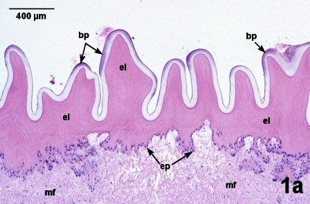
Figure 1a. The siphon of an adult geoduck clam has a thin layer of cuboidal to squamosal epithelial cells (ep) that separates the underlying muscular tissue (mf) from the two layers of acellular periostracum, a thick eosinophilic layer (el) and a lightly basophilic outer layer with occasional irregular darker basophilic patches (bp).

Figure 1b. The muscular mantle just anterior to the siphon of a juvenile geoduck clam. As in the adult, the epithelial cells (ep) separate the underlying muscular tissue (mf) from the acellular periostracum. However, in the posterior third of the muscular mantle next to the siphon, the distal layer of the periostracum is modified to form thorn-shaped structures (th) on the surface of the eosinophilic layer (el).
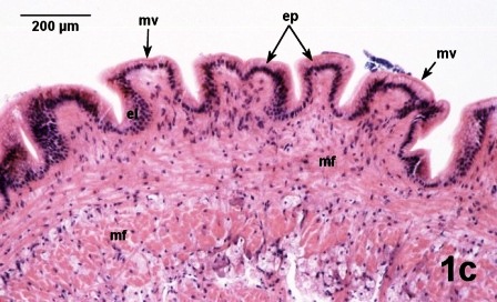
Figure 1c. The contracted siphon of a Pacific littleneck clam, Protothaca staminea, illustrates the underlying muscular tissue (mf) with an integument of columnar epithelial cells (ep) that have an outer surface of microvilli (mv) rather than a periostracum.

Figure 1d. At a higher magnification, the relaxed siphon of a Manila clam, Venerupis philippinarum. illustrates details of the outer surface of microvilli (mv) covering the columnar epithelial cells (ep) and the underlying muscular tissue (mf).
Mantle
The viscera are surrounded by the mantle cavity, which is defined by the mantle tissues. A thin mantle lies against the inner surface of the shell extending from the dorsal surface to the pallial line. It is constructed of loose vesicular connective tissue and a few scattered muscle fibres lying between a columnar epithelium on the distal surface (against the shell) (Fig. 2), and a simple cuboidal epithelium lining the mantle cavity. The columnar epithelium which covers the entire distal surface of the thin mantle has a brush border (or microvilli) on its surface.
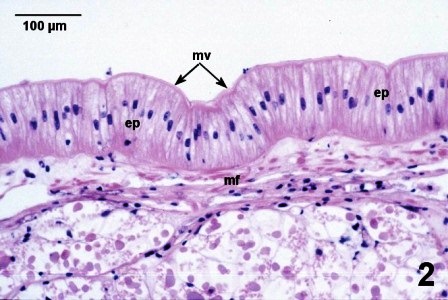
Figure 2. Cross section through the distal surface of the thin mantle that covers the inner surface of the shell. This tissue has relatively few muscle fibers (mf) and is covered by columnar epithelial cells (ep) with microvilli (mv) that are directed towards the shell. Note that the nuclei in the epithelial cells are located towards the centre of the cell and not basal as in the epithelial cells in the area of the glandular tissue (see Fig. 3a below).
A dark brown, thickened glandular area (Fig. 4 label bg on the Anatomy Page) of the thin mantle covers the dorsal ends of the left and right gills and labial palps and is ventral to the hinge and may function as a secretory gland. Histologically, this area has the appearance of an holocrine gland (Fig. 3a and b). Two, thin, eosinophilic, acellular layers lie between the shell and the simple columnar epithelial cells of this brown gland (Fig. 3a). The gland is composed of epithelial cells adjacent to each other which are sometimes arranged in follicles that are surrounded by muscle fibres interspersed with vesicular connective tissue cells. These epithelial cells contain a single, large vacuole full of a granular material that is brown-red when stained with haematoxylin and eosin. In some areas, the cells appear to have sloughed into a lumen, their nuclei becoming compact and pycnotic, and the contents of the vacuoles more dense and uniform (Fig. 3b).
Figures 3a and 3b. Glandular tissue in the mantle adjacent to the hinge of geoduck clams. Haematoxylin and eosin stain.
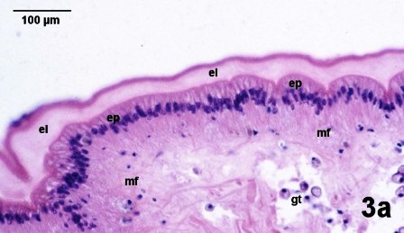
Figure 3a. The columnar eithelium (ep) is attached to the shell via two acellular eosinophilic layers (el) and there is a layer of muscular fibers (mf) between the epithelium and glandular cells (gt).

Figure 3b. A follicle of glandular tissue (gt) surrounded by muscle fibers (mf) and containing sloughed cells (sc) in the lumen.
The thick, fused muscular mantle forms the ventral and anterior surface of the mantle cavity, this surface is composed of ciliated simple columnar epithelium with goblet cells. Two raised pads of glandular tissue extend the length of the muscular mantle on either side of the midline (Fig. 4). A small (about 2x4 mm) conical papilla projects into the infrabranchial chamber at the dorsal end of the pedal aperture.
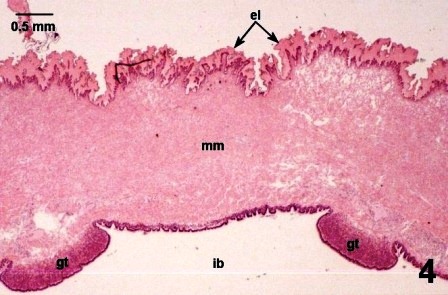
Figure 4. Cross section of the two raised pads of glandular tissue (gt) on the thick muscular mantle (mm) are oriented into the infrabranchial chamber (ib) of the mantle cavity of a juvenile geoduck clam. Note the acellular eosinophilic layer (el) on the outer surface of the muscular mantle. Haematoxylin and eosin stain.
References
Morse, M.P. and Zardus, J.D. 1997. Bivalva. Microscopic Anatomy of Invertebrates Vol. 6A Mollusca II. F.W. Harrison and A.J. Kohn. Wiley-Liss. pp. 7-118.
Simkiss, K. 1988. Molluscan Skin (excluding Cephalopods). The Mollusca Vol. 11 Form and Function. E.T. Truman and M.R. Clarke. Academic Press Inc. pp. 11 - 35.
Citation Information
Bower, S.M. and Blackbourn, J. (2003): Geoduck clam (Panopea generosa): Anatomy, Histology, Development, Pathology, Parasites and Symbionts: Normal Histology - Outer Surfaces.
Date last revised: August 2020
- Date modified: