
Geoduck clam (Panopea generosa): Anatomy, Histology, Development, Pathology, Parasites and Symbionts
Epibionts of the Geoduck clam
When cultured juvenile geoduck clams were maintained in suspension systems (e.g., pearl nets) for several months prior to being planted in the substrate, they acquired a superficial growth of diverse epibionts. The epibionts were attached to the surface of the integument covering the siphon and muscular mantle and on the shell. Although the relationship between the epibionts and their hosts was, in some instances, very close, no pathology was observed. Examples of histological sections through some of the various epibionts on the surface of juvenile geoduck clams (1995 year class) are presented in the following figures.

Figure 1. Various epibionts within the acellular periostracum on the siphon of a juvenile geoduck clam. Although the periostracum is disrupted (in comparison to unaffected specimens and Fig. 5 below), the underlying epithelium seems undamaged.

Figure 2a. Section through a polychaete worm (arrow), on the surface of the shell beside two bryzoans

Figure 2b. Section through a polychaete worm (arrow), on the surface of the siphon and surrounded by empty cuticles of a colonial bryzoan. The polycheate tube is fused to the periostracum but with no evidence of harm to the tissues of the juvenile geoduck clam.

Figure 3. Section through two bryzoans on the surface of the shell of a juvenile geoduck clam.

Figure 4. The test of a tunicate (arrow) is fused with the acellular periostracum of the siphon of a juvenile geoduck clam with no apparent harm to either organism.
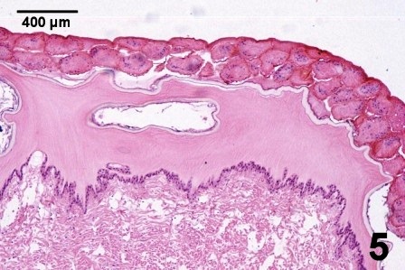
Figure 5. Numerous invertebrate eggs attached to the surface of the siphon of a juvenile geoduck clam. In this example, the structure of the acellular periostracum has not been altered and the underlying tissue looks unaffected.
All epibionts were lost soon after the juvenile geoduck clams (seed) were planted in the substrate. Although there was no apparent damage (histopathology) caused by the epibionts, growth data indicated that the epibionts appeared to be direct competitors with the juvenile geoduck clams for planktonic food. The procedure of holding geoduck clam seed in suspension culture for prolonged periods is no longer practiced.
References
Bower, S.M., J. Blackburn and G.R. Meyer. 1992. Parasite and symbiont fauna of Japanese littlenecks, Tapes philippinarum (Adams and Reeve, 1850) in British Columbia. Journal of Shellfish Research 11(1): 13-19.
Cheng, T.C. 1967. Marine molluscs as hosts for symbioses with a review of known parasites of commercially important species. In: F.S. Russell (ed.). Advances in Marine Biology. Volume 5. Academic Press Inc., London, p. 315-335.
Simkiss, K. 1988. Molluscan Skin (excluding Cephalopods). The Mollusca Vol. 11 Form and Function. E.T. Truman and M.R. Clarke. Academic Press Inc. pp. 11 - 35.
Citation Information
Bower, S.M. and Blackbourn, J. (2003): Geoduck clam (Panopea generosa): Anatomy, Histology, Development, Pathology, Parasites and Symbionts: Epibonts of the Geoduck Clam.
Date last revised: August 2020
- Date modified: