
Diagnostic Testing
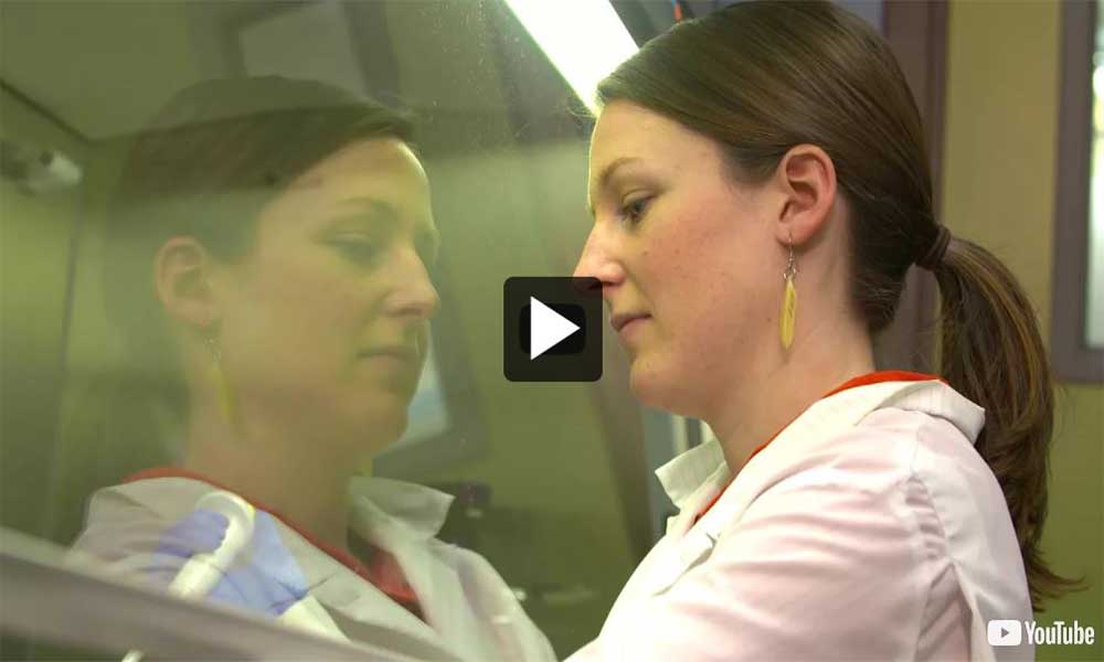
Video: A Day in the Life of a Fish Sample
Video: A Day in the Life of an Oyster Sample
Canada, as a member of the World Trade Organization (WTO), is obliged to implement an aquatic animal health program that meets the World Organization for Animal Health (OIE) standards recognized by the WTO. According to the OIE, “to ensure confidence in test results, both nationally and internationally, test methods must be properly validated with respect to their intended application, and they must be performed under appropriate control by proficient laboratory analysis. Confidence in test results requires that laboratories operate to a recognized standard of quality”. To meet OIE international standards, DFO NAAHLS laboratories are developing a quality management system for quality, administrative, and technical operations under the ISO/IEC 17025:2005 guidelines for accreditation by the Standards Council of Canada (SCC), standards also recommended by the World Organization for Animal Health. Laboratory customers, regulatory authorities, and accreditation bodies use this standard to confirm or recognize the competence of laboratories. This involves the implementation of very strict sample acceptance criteria, chain of custody requirements (i.e., the ability to track a sample through the full diagnostic process - from the collection of the sample in the field to the final results), and the development of standard operating procedures. DFO’s National Aquatic Animal Health Laboratory are responsible for delivering sound diagnostic testing in support of aquatic animal health responsibilities under the Health of Animal Act’s NAAHP program, as well as the Fisheries Act.
Diagnostic testing is used to detect pathogens in samples from fish, shellfish, or crustaceans. The presence of a pathogen does not mean a disease is also present. Diagnostic test results need to be considered in a broader context with other information, such as clinical signs or mortality, to determine if disease is present within a population. The presence of disease can affect the ability of fish, shellfish or crustaceans to grow, reproduce, thrive, or survive. Test results are used to support regulations and make management decisions; therefore the tests must be suitable or validated for their intended purpose. Validation can be a very elaborate and extensive process. NAAHLS diagnostic testing is performed in a biosecure environment with a strong quality management framework and laboratory information management system providing thorough documentation and traceability.
Training of lab staff is a key component in the NAAHLS quality management system. All lab analysts conducting diagnostic tests for regulated pathogens (i.e., reportable, immediately, notifiable, and annually notifiable diseases) must be trained and must demonstrate that they are competent for each diagnostic test. If an analyst is not successful in demonstrating their competency for a diagnostic test for a specific pathogen, they are not permitted to conduct that test on diagnostic samples until competency is achieved. Training is an ongoing activity and lab analysts’ competency for diagnostic tests is reviewed yearly.
The presence of a pathogen does not mean a disease is present. Diagnostic test results need to be considered with other information, such as clinical signs of disease or mortality, to determine if a disease is active within a population.
Not all strains of a pathogen cause disease. For example, some pathogens may have strains that cause no clinical signs of disease in animals while other strains may cause significant disease in animals leading to mortalities.
As part of DFO’s NAAHLS quality management system, the competency of lab analysts to conduct diagnostic tests for regulatory pathogens is reviewed annually. If an analyst cannot successfully demonstrate their competency for a diagnostic test for a specific pathogen, they are not permitted to conduct that test on diagnostic samples until competency is achieved.
A Quality Management System for Valid Laboratory Results - ISO/IEC 17025:2005
A quality management system in veterinary testing laboratories is achieved by developing and implementing good management practices, valid test and calibration methods, proper analytical techniques, staff training, and quality control and quality assurance.
Diagnostic Test Development
New tests are continuously evaluated and developed to address the needs of the program, such as detection of a new pathogen/strain, detection of a pathogen in a new host/matrix, to develop test methods that are more sensitivity and/or economical, allow non-lethal sampling and improve the quality control in the laboratory. When considering the development of a test intended for diagnostic use, the NAAHLS follow OIE validation guidelines to make sure the test is fit for purpose and properly characterized. Continuous improvement and delivery of quality testing results are key philosophies in the NAAHLS quality management system. Developing and validating quality controlled diagnostic tests are critical to accurately detect and identify pathogens and allow managers to make informed decisions based on sound data and scientific advice.
Diagnostic Test Validation
Virus isolation, bacteriology, and histology have been used historically for the detection of viruses, bacteria, and parasites and are recommended in scientific literature for the detection of several pathogens. Since the launch of the NAAHP, the NAAHLS laboratories have initiated test method validation, with a particular emphasis on molecular methods such as qPCR. Validation is a process that determines the fitness of a test method, which has been properly developed, optimised and standardised, for its intended purpose. Validation includes, but is not limited to, accuracy, precision, specificity, detection limit, limit of quantitation, linearity, range, and ruggedness or robustness. Validation is an on-going process that assesses a test method’s performance and characterizes it. To maintain a validated assay status, however, it is necessary to carefully monitor the assay’s performance over time under conditions of routine use to assure it maintains its fitness for purpose. DFO NAAHLS validation processes are based on the OIE’s validation guidelines.
Sample Processing
All submissions that arrive at the laboratory are assessed to determine if they are fit for testing and assigned a unique identifier or case number and relevant submission information is recorded. The case number follows the sample and all subsamples to ensure traceability from receipt to processing, testing, reporting and disposal. If required, whole fish, shellfish, and crustaceans are necropsied and relevant tissue or organ samples are collected for testing. Once samples are tested, a report is prepared and results are sent back to the customer. During any stage of the process, detection of a reportable pathogen is immediately reported to the Canadian Food Inspection Agency. Once testing is completed and the final reports sent, samples are archived or disposed of appropriately in a manner consistent with the NAAHLS’s policy on sample retention.
The NAAHLS laboratories have a wide range of diagnostic tools that are used in the detection of pathogens. The tests that are most frequently used in our laboratories are necropsy (gross observation), virus isolation, bacterial culture, histology, and polymerase chain reaction (PCR). Several other tests are also available, but are not as broadly used.
Samples of animals collected for diagnostic testing, such as tissues or organs, must follow strict shipping standards to ensure that samples arrive at the lab in a condition suitable for diagnostic testing. Otherwise samples that have not been properly stored degrade or decompose and diagnostic test results may not be accurate.
Pathogen Detection Techniques
-
False Positives / False Negatives
False Positives / False Negatives
The critical step in preventing and controlling any aquatic animal disease lies in the ability to accurately detect and identify the pathogen causing the disease. A false negative is a test result that incorrectly classifies an animal as free from exposure to or infection with a pathogen when in fact the animal is infected with the pathogen (often related to the pathogen being present at extremely low levels). A false positive is a test result that incorrectly classifies an animal as exposed to or infected with a pathogen when the animal is not infected with the pathogen.
During testing, steps are taken to avoid any false positive or false negative test results. A detailed protocol describing the steps of the test method is followed every time the test is performed. During the testing process, the use of appropriate positive and negative controls and thorough record keeping as per laboratory quality and information management systems ensures the validity of the test results. In addition, all DFO analysts undergo thorough training and must be authorised for specific testing processes before they are allowed to undertake any testing of diagnostic samples. When a positive result is obtained using one test, it is common practice in a diagnostic lab to use other test methods to confirm the result of an initial test. When results are used to make regulatory management decisions, it is important to have confidence in those results and to understand exactly what they represent.
- A false positive test result
- identifies an animal as infected with a pathogen when in fact it is free of the pathogen.
- A false negative test result
- identifies an animal as free of a pathogen when in fact it is infected with the pathogen.
-
Necropsy
Necropsy
When whole animals are received at the laboratory, the exterior of the animal and the internal organs are inspected for signs of disease. Any anomalies, for example lesions involving tissue damage, erosions or ulcers, discolouration, hemorrhage or swelling are recorded as these can help in determining if pathogens are present and what impact they might have. During necropsy, pieces of tissue and organs are harvested for further analysis using other diagnostic tests.
-
Virus Isolation
Virus Isolation
Just like humans and other animals, aquatic animals are prone to viral infections. Viruses are obligate intracellular parasites that require living cells in order to replicate. Although embryonated eggs and laboratory animals are very useful for the isolation of certain viruses, cell cultures are the sole system for virus isolation in most aquatic animal health laboratories. To prepare a sample for testing by virus isolation test method, tissues harvested during necropsy are prepared and processed in order to obtain a homogeneous mixture, which is then inoculated on specific cell lines and then incubated and monitored for a specific length of time and temperature appropriate for the virus. If a virus is present in the sample, it will replicate (i.e., increase in number) and eventually cause changes and damage to the cells. The changes and damage, collectively called cytopathic effect (CPE), are visible under the microscope. It can take several days or weeks for certain fish viruses to cause visible changes in the cells, so analysts need to observe inoculated cell cultures every few days. If CPE is observed, additional steps are taken to confirm the identification of the virus.
-
Bacteriology
Bacteriology
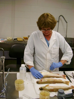
A DFO analyst inspects and collects tissues from whole fish submitted to be tested for bacterial and viral pathogens.
Source: Fisheries and Oceans Canada.Bacteriology is a branch of microbiology that studies bacteria, which are single cell organisms that can cause disease in aquatic animals. In order to test for bacterial pathogens during necropsy, a small amount of the target tissue is collected and spread with an inoculating loop on a media surface in a petri-dish. These petri-dishes are then incubated at a suitable temperature for the growth of bacteria for several days. Media in petri dishes contain a gel-like substance containing nutrients required for bacterial growth. If bacteria are present in the tissue, the bacteria will feed on the nutrients and reproduce to make bacterial colonies. If bacterial colonies are observed on the media further testing is conducted to identify them.
-
Histology
Histology
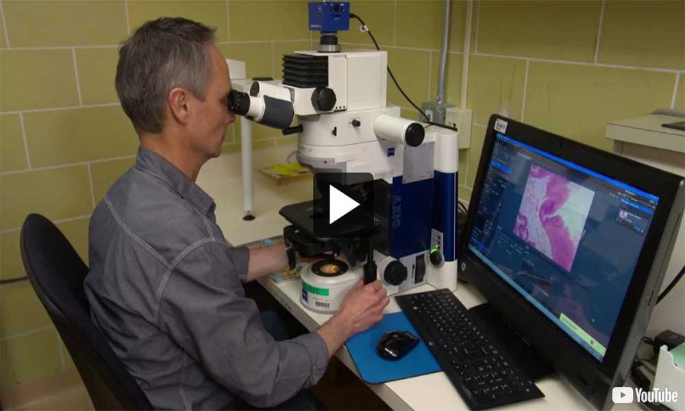
Histology is the observation of tissues and cells under the microscope. Although histology is used to identify specific pathologies in fish and crustaceans, it is the most widely used tool for detecting pathogens in molluscs. During necropsy, targeted tissues are collected, fixed in chemicals, and then processed so that the tissues are embedded in a paraffin block. Ultra-thin sections are then cut from paraffin blocks and placed on microscope slides. Once dried, the slides are stained using biochemical processes that color the tissues and then they are sealed. The most common stain used for shellfish histology is haematoxylin and eosin. The resulting slide is then observed on the microscope by an analyst. Analysis is performed following a specific protocol. The general physiological condition of the animal can be assessed and detection of the parasites is made by direct visual observation of the parasite in the tissues.
-
Molecular Biology
Molecular Biology
Every virus has a unique genetic fingerprint. The polymerase chain reaction is a technique widely used in a molecular laboratory to detect and identify specific portions of a virus’ genetic fingerprint. PCR testing provides an accurate diagnosis and is used in aquatic animal health diagnostic laboratories for the detection of nucleic acid (DNA or RNA) of bacteria, parasites, or viruses. In the NAAHLS laboratories this technique currently focuses on the detection of DNA or RNA target of one pathogen at a time. PCR tests are either used in its quantitative or conventional form. Quantitative PCR (qPCR) is highly sensitive and specific; data is collected in real time and allows a high throughput analysis. A positive reaction is detected by accumulation of fluorescent signal and results are expressed as a numerical value if a pathogen is present in a sample. The numerical value represents the cycle threshold (Ct) count, a Ct value is defined as number of cycles required for a fluorescent signal to cross the threshold or background signals. Ct values are inversely proportional to the amount of pathogen present in the sample i.e., the lower the Ct value the greater the amount of pathogen in a test sample as fewer cycles would be required to detect the pathogen. In a conventional PCR, PCR product is visualised on a gel. The distance to which a band migrates determines if a targeted pathogen is detected. Although the conventional PCR does not allow a high throughput of samples, the product of conventional PCR is used to obtain the genetic sequence of a detected pathogen.
Detection of a short genetic target of viral RNA belonging to a pathogen by PCR constitutes a presumptive positive test result. Due to high sensitivity of PCR assay, PCR test has a potential to produce false positive results and may require further testing to confirm these results. This is known as confirmatory testing.
Confirmatory testing may be done in two steps. The first step is to attempt to isolate the virus from fish tissues using cell culture. Cell culture is a specific medium that allows the virus to infect the cultured cells and multiply as it would in the host fish. It is possible to have a positive PCR test, but a negative cell culture result because cell culture requires a minimum dose of live virus to be present in the test sample to produce a positive result. Depending upon the virus, cell culture test can take up to four weeks to produce the results. The second step involves identifying the virus, which usually requires conventional PCR techniques to amplify longer and different portions of viral genes which are then sequenced and compared to a known fingerprint unique for that virus.
-
Tissue Quality
Tissue Quality
There are several factors which must be considered about the quality of the samples/fish tissue before reliable testing by PCR can occur. Since PCR requires samples to be either fresh or appropriately preserved, fish should be collected live, moribund (i.e., dying), or as fresh mortalities (within 24 hours). Since both the host (fish) and viral RNA degrade rapidly after death, virus detection can quickly become challenging by PCR or any other accepted test methods. Since RNA degrades rapidly an extra test called the "reference gene assay", may be conducted to determine the quality of the original tissue. The results of this procedure will indicate the level of decomposition of the fish sample by comparing it to a well preserved sample. If the RNA has substantially degraded, neither a PCR nor any other approved test method will accurately determine the presence or absence of the virus with any degree of confidence. If there are concerns regarding the degradation of the sample, other tests can be conducted to verify whether the test results are accurate.
Fish tissue collected for PCR analysis should be either frozen immediately or stored in ‘RNA later’ in order to preserve the RNA. Since tissue and virus degradation occurs at -20°C, samples should be stored at -70°C for proper preservation.
Secondly, since virus targets specific organs, it is important to collect the appropriate organ/tissue for PCR testing. Gills and/or other external organ tissue can also be tested; however, results should be treated with caution since the detection may indicate the presence of viral particles in the environment and not necessarily indicate that the fish is infected with the virus.
Finally, the size of the organ or tissue sampled should be large enough for testing. A tissue sample the size of a grain of rice can be tested for both PCR and molecular confirmatory tests (gene sequencing); however, significantly larger amounts are needed for cell culture and archiving for future reference and testing.
- Date modified: