
Marteilioides sp. of Clams
On this page
Category
Category 1 (Not Reported in Canada)
Common, generally accepted names of the organism or disease agent
Marteilioides of clam oocytes.
Scientific name or taxonomic affiliation
Marteilioides sp. in the phylum Paramyxea as proposed by Desportes and Perkins (1990) and supported by Berthe et al. (2000). The morphology of this parasite was similar to that of Marteilioides chungmuensis in the gonad of oysters from Korea and Japan but the relationship to this parasite is not known.
Geographic distribution
Korea and Japan.
Host species
Venerupis (= Tapes) philippinarum.
Impact on the host
A Marteilioides- like parasite was reported from 1.6% of about 2600 V. philippinarum from the Hadong and Namhae coasts of southern Kyongnam province, Korea between March 1996 and April 1997 (Lee et al. 2001). A similar parasite was also detected in September 2003 in the oocytes of one of 40 clams experimentally deployed from Oita Prefecture to Yamaguchi Prefecture, Japan in April 2002 to investigate the drastic decrease in commercial V. philippinarum stocks (Itoh et al. 2005). In all cases, infection did not cause any significant host reaction.
Diagnostic techniques
Histology
From one to eight round plasmodia (stem cells) containing two sporonts (secondary cells) each with one spore (tertiary cell), resulting from exogenous budding, occurred within the cytoplasm of oocytes and ova of infected clams.

Figure 1. Ovarian follicles of Venerupis philippinarum from Japan containing ova infected with Marteiloides sp. (arrows).
Electron Microscopy
The mature spore consisted of one outer cell and two inner cells which contained haplosporosomes (Itoh et al. 2005).

Figure 2. Plasmodium (C1) of Marteiloides sp in an ova of Venerupis philippinarum from Japan containing two sporonts (secondary cells, C2), each with a developing spore (tertiary cell, C3).
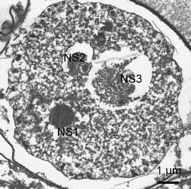
Figure 3. Spore of the Marteiloides sp. in Fig. 2 consisting of three cells produced by internal cleavage (NS1, nucleus of spore cell; NS2, nucleus of secondary cell; NS3, nucleus of tertiary cell) and haplosporosomes (H).
Methods of control
No known methods of prevention or control. Infected clams should not be transported into areas known to be free of the disease.
References
Berthe, F.C.J., F. Le Roux, E. Peyretaillade, P. Peyret, D. Rodriguez, M. Gouy and C.P. Vivares. 2000. Phylogenetic analysis of the small subunit ribosomal RNA of Marteilia refringens validates the existence of Phylum Paramyxea (Desportes and Perkins, 1990). The Journal of Eukaryotic Microbiology 47: 288-293.
Desportes, I. and F.O. Perkins. 1990. Phylum Paramyxea. In: Margulis, L., J.O. Corliss, M. Melkonian and D.J. Chapman (eds), Handbook of Protoctista. Jones and Bartlett Publishers, Boston, MA. Pg. 30-35.
Itoh, N., K. Momoyama and K. Ogawa. 2005. First report of three protozoan parasites (a haplosporidian, Marteilia sp. and Marteilioides sp.) from the Manila clam, Venerupis (= Ruditapes) philippinarum in Japan. Journal of Invertebrate Pathology 88: 201-206.
Lee, M.-K., B.-Y. Cho, S.-J. Lee, J.-Y. Kang, H.D. Jeong, S.H. Huh and M.-D. Huh. 2001. Histopathologucal lesions of Manila clam, Tapes philippinarum, from Hadong and Namhae coastal areas of Korea. Aquaculture 201: 199-209.
Citation information
Bower, S.M., Itoh, N. (2005): Synopsis of Infectious Diseases and Parasites of Commercially Exploited Shellfish: Marteilioides sp. of Clams.
Date last revised: June 2005
- Date modified: