
Malpeque Disease of Oysters
On this page
Category
Category 2 (In Canada and of Regional Concern)
Common, generally accepted names of the organism or disease agent
Malpeque disease.
Scientific name or taxonomic affiliation
Unknown aetiology.
Geographic distribution
Atlantic Canada (with the exception of Bras d'Or Lakes, Cape Breton and an isolated low salinity population in southwest Nova Scotia).
Host species
Crassostrea virginica.
Impact on the host
Previously unexposed populations have historically demonstrated up to 99% cumulative mortality, with survivors showing apparent resistance. Low salinity appears to retard the disease, hence relict populations of oysters survive in upper estuaries of certain rivers within the Gulf of St. Lawrence. Infected animals show gross signs of mantle regression, gaping, oedema and abscesses in the mantle. Yellow-green scars were also observed on the inner surface of the shell. The agent causing this disease has yet to be clearly identified, however, it is highly infectious. Experimental transplants of "susceptible" stocks from disease-free Cape Breton to Malpeque Bay, in the late 1960s and in 1994, resulted in 90% mortality within 24 months. No epizootics associated with the disease have been reported since the 1950s (when the disease spread from Prince Edward Island to New Brunswick). It is assumed that all remaining C. virginica in the southern Gulf of St. Lawrence are resistant to the aetiologic agent of Malpeque disease
Diagnostic techniques
Gross Observations
Mantle regression, gaping, oedema and cream/yellow coloured abscesses up to 1 cm in diameter in the body wall. Yellow-green scars were also observed on the inner surface of the shell adjacent to lesions in the soft tissues.
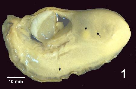
Figure 1. Crassostrea virginica removed from shell and illustrating lesions (arrows) typical of Malpeque disease
Histology
Oedema of the mantle; retardation of gonad development; connective tissue abscess lesions in conjunction with focal infiltration of haemocytes, especially in association with intestinal walls; and presence of large amounts of ceroid in some oysters, especially within the digestive gland. Presence of abnormal haemocytes characterized by enlarged nuclei and reduced cytoplasm. These features resemble haemic neoplasia in oysters but lack evidence of mitosis.

Figure 2. Histological section through part of the body (mainly digestive gland and adjacent intestinal tract) of Crassostrea virginica at an early stage of Malpeque disease showing focal patches of infiltrating haemocytes (IH) and retardation of gonadal development (RG)

Figure 3. Histological section through part of the body of Crassostrea virginica at an intermediate stage of Malpeque disease showing extensive patches of infiltrating haemocytes some of which surround small abscess lesions (Arrows) in the connective tissue among the digestive gland tubules.

Figure 4. Histological section through part of the body of Crassostrea virginica at the late stage of Malpeque disease showing large abscess lesions (arrows) in the digestive gland. The extensive abscess lesion (AL) adjacent to the intestinal tract replaced much of the digestive gland in this section of the oyster.
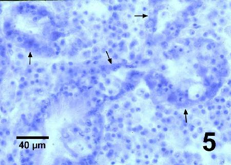
Figure 5. Higher magnification of Fig. 4 illustrating remnants of the digestive gland tubules (arrows) that are being infiltrated by numerous haemocytes.
Methods of control
Do not introduce oysters from areas which have been affected, historically, by Malpeque disease.
References
Drinnan, R.E. and J.C. Medcof. 1961. Progress in rehabilitating disease affected oyster stocks. Fisheries Research Board of Canada. General Series Circular No. 34, 3 pp.
Needler, A.W.H. and R.R. Logie. 1947. Serious mortalities in Prince Edward Island oysters caused by a contagious disease. Transactions of the Royal Society of Canada XLI(III) Section V: 73-89.
Li, M.F., C. Flemming and J.E. Stewart. 1967. Serological differences between two populations of oysters (Crassostrea virginica) from the Atlantic coast of Canada. Journal of the Fisheries Research Board Canada 24(2): 443-445.
Li, M.F., G.S. Traxler, S. Clyburne and J.E. Stewart. 1980. Malpeque disease: isolation and morphology of a Labyrinthomyxa-like organism from diseased oysters. International Council for Exploration of the Sea C.M.1980/F:15: 1-9.
Citation Information
McGladdery, S.E. and Bower, S.M. (1999): Synopsis of Infectious Diseases and Parasites of Commercially Exploited Shellfish: Malpeque Disease of Oysters.
Date last revised: October 1999
Comments to Susan Bower
- Date modified: